Another Confirmation Of Self Assembly Nanotechnology In COVID Bioweapons: Remarkable Longitudinal Study And Culture Work Of COVID Shots For Up To 12 Months And Cellular Toxicity Studies
Another Confirmation Of Self Assembly Nanotechnology In COVID Bioweapons: Remarkable Longitudinal Study And Culture Work Of COVID Shots For Up To 12 Months And Cellular Toxicity Studies
BREAKING: COVID SHOTS INSTALLED NANOBOTS
New Japanese study proves Pfizer and Moderna v*ccines contain unauthorized “animated worm-like” entities, invisible to the human eye, which swim, wiggle, and assemble themselves into complex structures, which cause clots inside the body. (What embalmer Richard Hirschman found and exposed in the film Died Suddenly).
Dr. Young Mi Lee and Dr. Daniel Broudy from Okinawa Christian University discovered these “undisclosed additional engineered components” by isolating unused vaxx vials for 3 weeks, and then examined them under 400X magnification.
Lee and Broudy saw that when the nanotechnology was energized it created “discs, chains, spirals, tubes, and right-angle structures.”
The researchers, who published their findings in the International Journal of Vaccine Theory Practice and Research, believe these mysterious nanoparticles are responsible for the explosion of “turbo cancer” and autoimmune diseases around the globe.
They also concluded in the study their suspicion that these smart microscopic components are part of the elite’s “long-planned well-funded Internet of Bodies,” which was described as a kind of “synthetic global central nervous system” turning humans into controllable “Biohybrid Magnetic Robots.”
The study ends by calling for a global ban on all mRNA shots, until these nanobots are studied long-term. They also demanded the labels “v*ccine” and “safe and effective” be removed because the concoction injected into billions is officially neither.
Another Confirmation Of Self Assembly Nanotechnology In COVID Bioweapons: Remarkable Longitudinal Study And Culture Work Of COVID Shots For Up To 12 Months And Cellular Toxicity Studies
In this important study, Dr Young Mi Lee with Dr Daniel Broudy describes her culture findings of Pfizer and Moderna Bioweapons. Many different studies were done, including incubation of the COVID injections, cellular effects upon blood and semen specimen. The tests showed direct toxicity to semen and death of all sperm within minutes up to 1.5 hours in the healthiest donor samples when put in contact with the “vaccine” solution. EMF exposure was also tested, in addition with different supplements and treatment solutions. For this substack, select images were taken from the article which is 66 pages long. Please refer to this link and download the paper - please share widely - this is one the most comprehensive and long term study of the COVID injections and their cellular effects to date. The findings are entirely consistent with all prior research findings of self assembly nanotechnology from the injectables.
Real-Time Self-Assembly of Stereomicroscopically Visible Artificial Constructions in Incubated Specimens of mRNA Products Mainly from Pfizer and Moderna: A Comprehensive Longitudinal Study
Abstract
Observable real-time injuries at the cellular level in recipients of the “safe and effective” COVID-19 injectables are documented here for the first time with the presentation of a comprehensive description and analysis of observed phenomena. The global administration of these often-mandated products from late 2020 triggered a plethora of independent research studies of the modified RNA injectable gene therapies, most notably those manufactured by Pfizer and Moderna. Analyses reported here consist of precise laboratory “bench science” aiming to understand why serious debilitating, prolonged injuries (and many deaths) occurred increasingly without any measurable protective effect from the aggressively, marketed products. The contents of COVID-19 injectables were examined under a stereomicroscope at up to 400X magnification. Carefully preserved specimens were cultured in a range of distinct media to observe immediate and long-term cause-and-effect relationships between the injectables and living cells under carefully controlled conditions. From such research, reasonable inferences can be drawn about observed injuries worldwide that have occurred since the injectables were pressed upon billions of individuals. In addition to cellular toxicity, our findings reveal numerous — on the order of 3~4 x 10⁶ per milliliter of the injectable — visible artificial self-assembling entities ranging from about 1 to 100 µm, or greater, of many different shapes. There were animated worm-like entities, discs, chains, spirals, tubes, right-angle structures containing other artificial entities within them, and so forth. All these are exceedingly beyond any expected and acceptable levels of contamination of the COVID-19 injectables, and incubation studies revealed the progressive self-assembly of many artifactual structures. As time progressed during incubation, simple one- and two-dimensional structures over two or three weeks became more complex in shape and size developing into stereoscopically visible entities in three-dimensions. They resembled carbon nanotube filaments, ribbons, and tapes, some appearing as transparent, thin, flat membranes, and others as three-dimensional spirals, and beaded chains. Some of these seemed to appear and then disappear over time. Our observations suggest the presence of some kind of nanotechnology in the COVID-19 injectables.
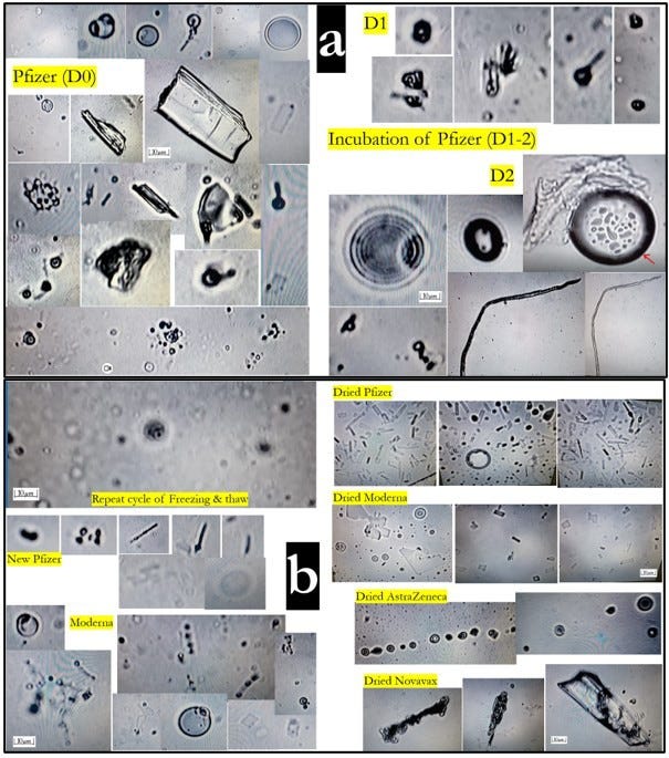
Figure 5. Direct microscopic findings observed in two dimensions magnified 400X: (a) Remnants and new Pfizer injectables, directly observed as well as after incubation for 1 -2 days. (b) Moderna and 4 dried COVID-19 injectables (Pfizer, Moderna, AstraZeneca, and Novavax).
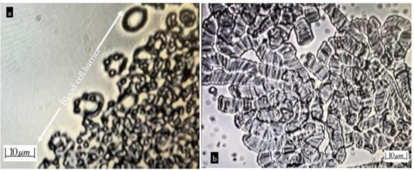
Figure 6. Interactions observed for whole blood(a)/plasma(b) with Novavax at 400X magnification: (a) Within 1 hour, blood cells formed a prominent barrier against “vaccine” contents. (b) After 30 minutes, severe aggregates of red blood cells in rouleaux appeared in the plasma specimen.
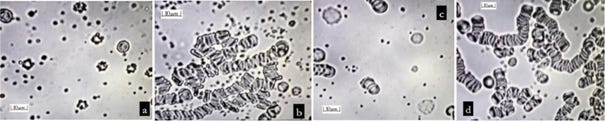
Figure 7. Plasma reactions after two hours with four COVID-19 injectables — Pfizer, Moderna, Novavax, and AstraZeneca: (a) Pfizer showing cellular collapse (pyknosis) of white blood cells and damaged platelets; (b) Moderna with stacks of red blood cells (rouleaux); (c) Novavax with the nucleus of white blood cells disintegrating (karyorrhexis), abnormal platelet aggregations, and some rouleaux of red blood cells; and (d) AstraZeneca with prominent Rouleau of red blood cells.
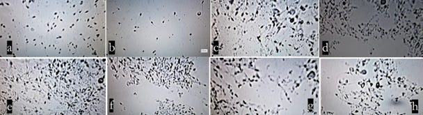
Figure 8. Reaction of semen to COVID-19 injectables at 200X magnification: (a) semen with normal saline as a control added after two hours; (b) with flu vaccine added as a control after 1.5 hours showed sperm cells with intact morphology and with typical progressive natural reduction in sperm motility; (c) 30 minutes after Pfizer-1 injectable was added, sperm motility showed rapid reduction; (d) Pfizer-1 after one hour, all sperm motility ceased;
(e) 30 minutes after Moderna. injectable added; (f) one hour after Moderna was added sperm cells were completely immotile; (g) 30 minutes after Novavax was added; (h) one hour after Novavax, all motility ceased.
Figure 11. Findings for Pfizer incubation study for 372 days; (a) Day 22, this is what we describe as a beaded chain (at 400X magnification); (b) Day 24, 2- dimensional geometric self-assembly at the bottom (at 200X magnification) in normal saline; (c) Day 60, floating 3-dimensional detailed chip-like structures (at 400X magnification) in distilled water; (d) and (e) day 60, accumulated 3-dimensional chip-like structures within an oval shaped boundary (200X/400X) in distilled water; (f), (g), (h), (i) Floating filaments shedding bubbles inside and outside in normal solution at day 95 (100x/100x/200x/200x); (j), (k), (l), (m) Progressive degenerative changes in distilled water 200X (day 82/day 256/day 306/day 372).
Figure 12. Various coils, ribbons, and spirals in Pfizer, distilled water: (a) Day 60 (at 200Xmagnification); (b) ~ (e) Day 74 (at 200X magnification); (f) Day 176 (at 100X magnification).
Figure 13. Beaded Chains and Assorted Structures in Pfizer Distilled Water (Day 176, 400x): (a) Various artificial satellite-like structures, (b) Long beaded chains gathered on the central surface of the medium.
Figure 14. Typical Algae-typed Magnetic Nanobot-like Spirals in Pfizer in distilled water: (a) Day 176 (400x); (b) Day 337 (200x).
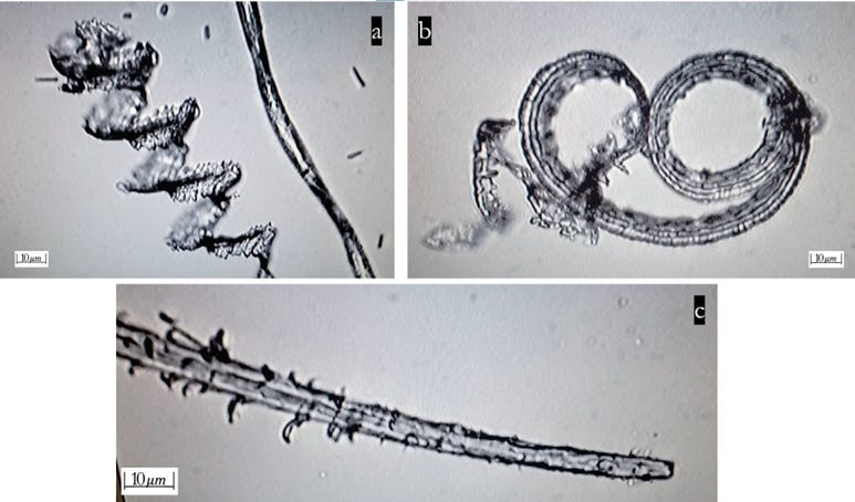
Figure 15. Various filaments — striated ribbons, sprouting in the late stage (Day 316) of incubation of Pfizer in distilled water: (a) and (b) curled striated ribbons (100x); (c) sprouting filaments in Pfizer (200x).
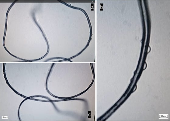
Figure 16. Bundle of transparent thin wire-like tubes with shedding bubbles in Pfizer incubation in distilled water (Day 331); floating in the uppermost layer (a- 40x/b-100x/c-40x).
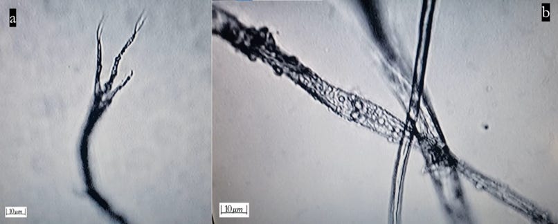
Figure 17. Tripod-like, striated filaments in Pfizer incubation in normal saline (Day 346, 200x): (a) More developed tripod-like structures or (b) Striated patterns on filaments.
Figure18. Curled Striated Ribbons and Bubbles in Pfizer Incubation in Distilled Water (Day 406 and 499); (a), (b), and (c) uniquely striated curled Ribbons in Pfizer in DW at 406 days of incubation (40X/100X/200X); (d), and (e); Bubbles (arrows) appeared on the surface of the curled ribbons at 499 days incubation (200X).
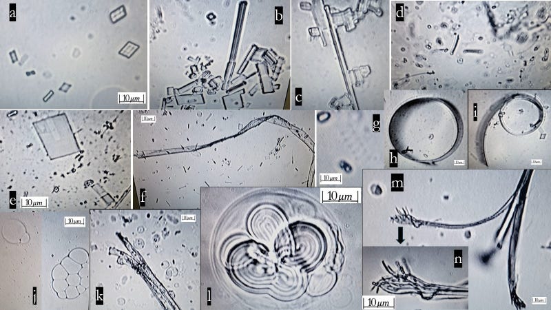
Figure 19. Geometric chip-like assembly, filaments, spirals, ribbons, and encapsulated wire bundles in the Moderna incubation study throughout Day 630 (100~400X): (a) Day 16; (b) Day 40; (c) Day 42; (d) Day 125; and (e) Day 126 (all 400x) in normal saline. (f) Day 126, chips and filament (100x) in normal saline; (g) Day 36, rarely observed small spring in distilled water; (h) and (i) Day 42, small circular ribbons in normal saline, (400x/200x); (j) Day 295, lobulated bubbles floating at the uppermost layer(100x) in distilled water; (k) Day 313, split-ended tape (200x) in distilled water; (l) Day 313, capsuled, well-packed wire bundles (400x) in distilled water; (m) Day 630, split-ended filament in normal saline (100x); (n) Day 630, magnified split-ended filament (400x).
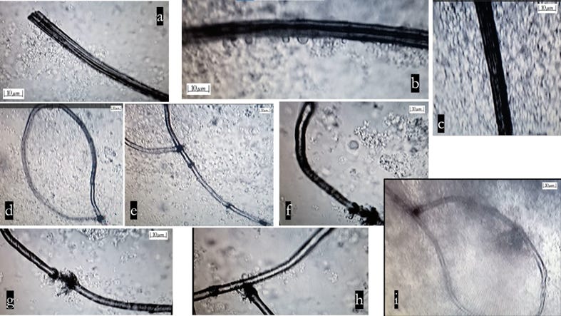
Figure 21. Findings of Moderna incubation study in plasma 2: (a) Day 133- dark pipe-like structure developed (100x); (b) with shedding bubbles (200x);
(c) disappeared bubbles at Day 282(100x); (d) and (e) Day 133, snare-like lasso tubes (100x); (f), (g), and (h) Day 133, broken, disconnected points in the lasso- like tube (200x); (i.) Day 282, still the same figure maintained (100x).
Figure 22. Heat (Warming) Study of Pfizer - after 48 hours of warming at BT in normal saline: (a), (b), (c), (d), and (e): Floating well-assembled 3-dimensional geometric structures on the surface of the media after 48 hours of warming (36.5°C) on the heat template. Accelerated development was similarly matched to the findings in the 2nd~ 3rd week of the unexposed incubation study (400x).
Figure 25. EMF (external hard drive/wireless recharger) study of Pfizer in distilled water (200X), 101 days of incubation: Pfizer study of exposure to external hard drive for 2 hours at Day 101 incubation in distilled water, and then followed by exposure to a wireless recharger for 2 hours. (a) after 2 hours of exposure to an external hard drive – more blunt edges and blurry alignment, more degraded than natural degeneration change; (b) after 2 hours of exposure to the wireless recharger immediately following exposure to the external hard drive- mild recovery (rescuing effect) appeared.
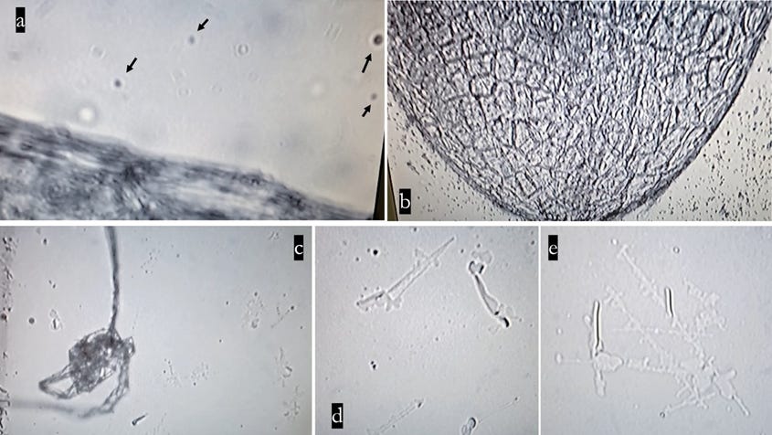
Figure 26. Recycling Pattern Study — Presumptive recycling pattern from the Moderna vaccinee’s skin extract (E1) in normal saline: (a) seed-like tiny dark particles (arrows) floating around the skin extract 1(E1) in the normal saline media (400x); (b) tiny particles scattered around the vaccinee’s skin extract, dark large material-crocodile skin-like structure (100x); (c) E1 seeds culture in normal saline - trace of the self-assembly at the bottom and floating filaments together after Day 366 incubation (100x); (d) and (e) Trace of the self-assembled geometric structures at Day 366 incubation (400x).
Source: Seemorerocks


Comments
Post a Comment